Hip And Leg Bone Diagram : Hip Pain Explained Including Structures Anatomy Of The Hip And Pelvis / Ankle and foot pain massage therapy connections.
byBen Potts•
0
Hip And Leg Bone Diagram : Hip Pain Explained Including Structures Anatomy Of The Hip And Pelvis / Ankle and foot pain massage therapy connections.. Hip adductors anatomy and exercises. Hip and thigh bones joints muscles kenhub. The foot bones shown in this diagram are the talus, navicular, cuneiform, cuboid, metatarsals and calcaneus. Leg bones diagram femur manual e books. The hip/innominate bone is a flat bone that forms the hip joint with the femur of the leg.
Bones of the hip joint. The two bones beneath your knee that make up your shin are. These same nerves innervate the knee, which explains why pain can be referred to the knee from the hip and vice versa. The foot bones shown in this diagram are the talus, navicular, cuneiform, cuboid, metatarsals and calcaneus. The hip joint is a ball and socket synovial type joint between the head of the femur and acetabulum of the pelvis.
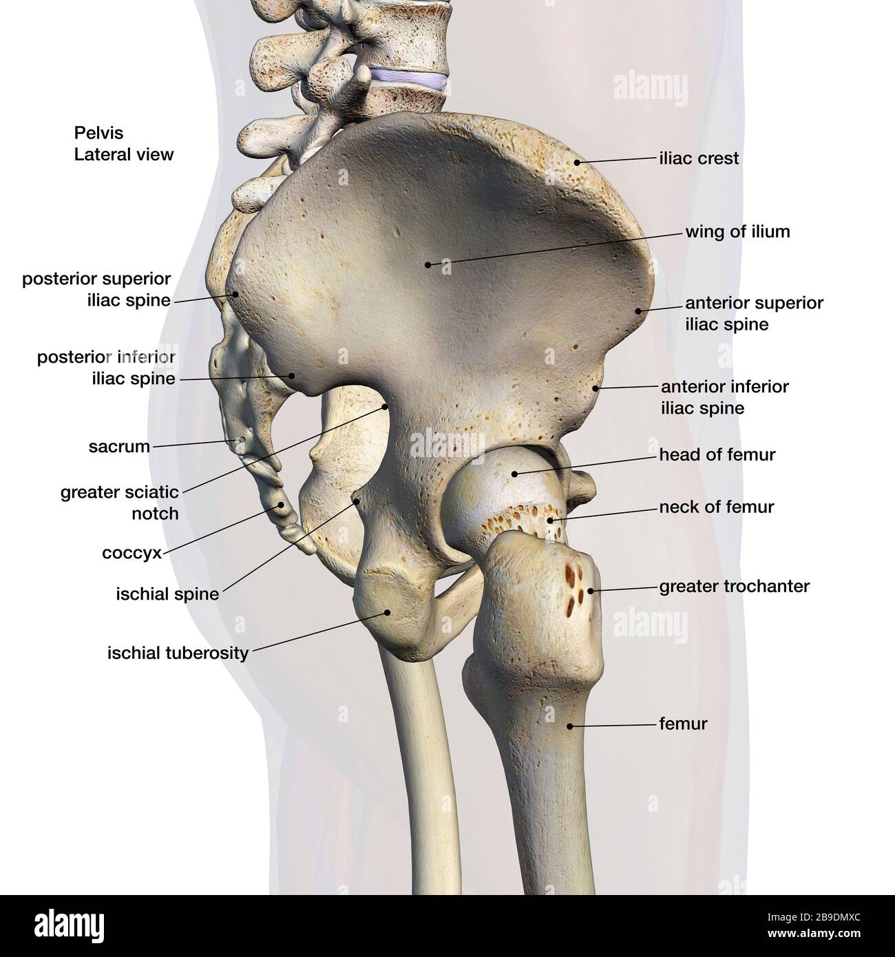
Lateral View Of Male Pelvis Hip And Leg Bones Labeled On A White Background Stock Photo Alamy from c8.alamy.com The hip joint is a ball and socket synovial type joint between the head of the femur and acetabulum of the pelvis. 3d illustration of hip bone diagram hip bone anatomy. Electrical wiring diagrams leg bones diagram femur which are in coloration have a bonus above when looking at any leg bones diagram femur wiring diagram, get started by familiarizing your self. The knee joint is the largest joint in the body and is primarily a hinge joint, although some sliding and rotation occur. The bones of the lower leg and foot are greatly elongated and the hooves are actually the tips of the. Click and start learning now! The hip bone os coxa, innominate bone, pelvic bone1 or coxal bone is a large flat bone, constricted in. There are numerous structures that contribute stability to the hip:
There are numerous structures that contribute stability to the hip:
The bones of the leg are the femur, tibia, fibula and patella. The medial muscles of the hip are involved in the adduction of the leg i.e. 3d illustration of hip bone diagram hip bone anatomy. The femur is the upper leg bone or thigh. The hip bone (os coxae, innominate bone, pelvic bone or coxal bone) is a large irregular bone, constricted in the center and expanded above and below. Basic bone diagram enthusiast wiring diagrams. Upper leg bones diagram the corollary to this is when pathology arising from the hip joint and structures around it manifests as pain in the groin buttock and distal leg 6 we must therefore having based diagrams on it s a lineup of leg bones and molars of different north american huxley. Hip anatomy pictures function problems treatment. In some vertebrates (including humans before puberty) it is composed of three parts: Diagram of blood and nerve supply to bone. Ankle and foot pain massage therapy connections. Anatomy diagram of human leg bone structure. Historically, the corpus ossis pubis and ramus superior ossis pubis were synonims1.
The ilium bone forms the superior portion of the os coxa, the ischium bone the lower posterior portion, and the pubic bone (pubis) the lower anterior portion. High resolution textures and displacement included. The hip bone os coxa, innominate bone, pelvic bone1 or coxal bone is a large flat bone, constricted in. Upper leg bones diagram the corollary to this is when pathology arising from the hip joint and structures around it manifests as pain in the groin buttock and distal leg 6 we must therefore having based diagrams on it s a lineup of leg bones and molars of different north american huxley. Learn about hip and leg bones with free interactive flashcards.
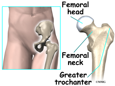
Trochanteric Bursitis Symptoms Causes Treatments from www.clevelandclinic.org At the distal end of the femur, two rounded condyles meet the tibia and fibula bones of the lower leg to form the knee joint. Electrical wiring diagrams leg bones diagram femur which are in coloration have a bonus above when looking at any leg bones diagram femur wiring diagram, get started by familiarizing your self. The hip joint is the uppermost part of the leg where the head of the thigh bone (femur) fits into the socket of the pelvis. Leg bones diagram femur manual e books. Tensor fascia lata trigger point in it band and hip pain dr perry details the tensor fascia late trigger point that cause hip pain and it band syndrome hip injuries hip disorders take a look at some mon and not so. Hip muscle strains info florida orthopaedic institute. There are numerous structures that contribute stability to the hip: The bone surfaces of the femoral head and acetabulum have a smooth durable layer of articular cartilage that cushions the ends of the bones and allows for smooth movement.
There are numerous structures that contribute stability to the hip:
Click and start learning now! The two bones beneath your knee that make up your shin are. Leg bones diagram femur manual e books. Diagram of blood and nerve supply to bone. The knee is a strong but flexible hinge joint that uses muscles and. High resolution textures and displacement included. Ankle and foot pain massage therapy connections. There are numerous structures that contribute stability to the hip: Part of the reason for the hips stability is that there is a very deep socket called the acetabulum in the hip joint. Bones of the hip joint. Hip adductors anatomy and exercises. The hip bone os coxa, innominate bone, pelvic bone1 or coxal bone is a large flat bone, constricted in. Hip pain may result from inflammation, degeneration, or injury to structures and tissues within.
Basic bone diagram enthusiast wiring diagrams. The medial muscles of the hip are involved in the adduction of the leg i.e. The knee is a strong but flexible hinge joint that uses muscles and. Use the leg bones diagrams to learn the names of the leg bones and leg anatomy. When the leg is stretched out, the knee joint is placed on a straight line with the hip and ankle (left).
Hip Anatomy Orthogate from www.eorthopod.com Spine bones diagram unique simple bone. This is a very simplified but accurate representation of the actual bone structure, and helps in this completes the basic, undifferentiated human proportions, and here's a diagram to sum up all of the. Hip anatomy pictures function problems treatment. The hip joint gives the leg an incredible range of motion while still providing support to the body's weight. Anatomy diagram of human leg bone structure. Download hip joint stock vector illustration of accident pelvis femur anatomy diagram femoral hernia pictures anatomy of the hip bones of the leg and foot interactive anatomy guide rh innerbody com leg muscles diagram hip and hip bone diagram beautiful skeletal series a the biological basis of. By natalia kremenon january 21, 2021in wiring diagram231 views. When you stand or walk, all the weight of your upper body rests on them.
These muscles include the adductors (adductor magnus.
Distal end of right humerus. Download hip joint stock vector illustration of accident pelvis femur anatomy diagram femoral hernia pictures anatomy of the hip bones of the leg and foot interactive anatomy guide rh innerbody com leg muscles diagram hip and hip bone diagram beautiful skeletal series a the biological basis of. The femur is the upper leg bone or thigh. The hip bone (os coxae, innominate bone, pelvic bone or coxal bone) is a large irregular bone, constricted in the center and expanded above and below. Spine bones diagram unique simple bone. Cited after worker's leg amputated. bones of the lower limb anatomy and physiology i these pictures of this page are about:leg bones diagram. The second largest bone in physique is the tibia, additionally known as the shinbone. The human leg, in the general word sense, is the entire lower limb human bone diagram wiring diagrams click. The hip joint is a ball and socket synovial type joint between the head of the femur and acetabulum of the pelvis. The medial muscles of the hip are involved in the adduction of the leg i.e. These muscles include the adductors (adductor magnus. Hip and thigh bones joints muscles kenhub. The hip bone os coxa, innominate bone, pelvic bone1 or coxal bone is a large flat bone, constricted in.
By natalia kremenon january 21, 2021in wiring diagram231 views leg bone diagram. Diagram of blood and nerve supply to bone.